Home
/ Anatomy Of Back Of Neck - Best Muscle Anatomy With Labels Stock Photos, Pictures ... : Anatomical principles underlying cranial nerve lesions;
Anatomy Of Back Of Neck - Best Muscle Anatomy With Labels Stock Photos, Pictures ... : Anatomical principles underlying cranial nerve lesions;
Anatomy Of Back Of Neck - Best Muscle Anatomy With Labels Stock Photos, Pictures ... : Anatomical principles underlying cranial nerve lesions;. Neck, in land vertebrates, the portion of the body joining the head to the shoulders and chest. In the neck, the platysma when contracted throws the skin into oblique ridges parallel with the fasciculi of the muscle. Watch cervical muscle anatomy animation. The physicians originally studying human anatomy thought the skull looked like an helmet. The back of the neck is mostly comprised of muscles, as well as the spine.
Dummies has always stood for taking on complex concepts and making them easy to understand. Muscle head anatomy vocal organ diagram female neck anatomy neck wireframe head neck human anatomy head artery anatomy face pharynx vector neck degree head anatomy 3d. Anatomical principles underlying cranial nerve lesions; Despite being a relatively small region, it contains a range of important anatomical features. The pll starts at c2 and goes down the back of the vertebral bodies and intervertebral discs.
Head and Neck Bones Full Color Vintage Print Atlas of Human from img1.etsystatic.com When to have lower back surgery. Dummies helps everyone be more knowledgeable and confident in applying what they know. Your neck is like no other part of the vertebral spinal column and enables your head and neck a wide range of motion. Watch cervical muscle anatomy animation. Demonstrate sound knowledge of the surface/living and radiological anatomy of the head, neck and. A collection of anatomy notes covering the key anatomy concepts that medical students need to learn. The word neck comes from a latin word which means cervical. Click now to study the muscles, glands and organs of the neck at kenhub!
3d video tutorials and interactive modules on the anatomy of the back including anatomy of the musculature, vertebral column, joints and ligaments.
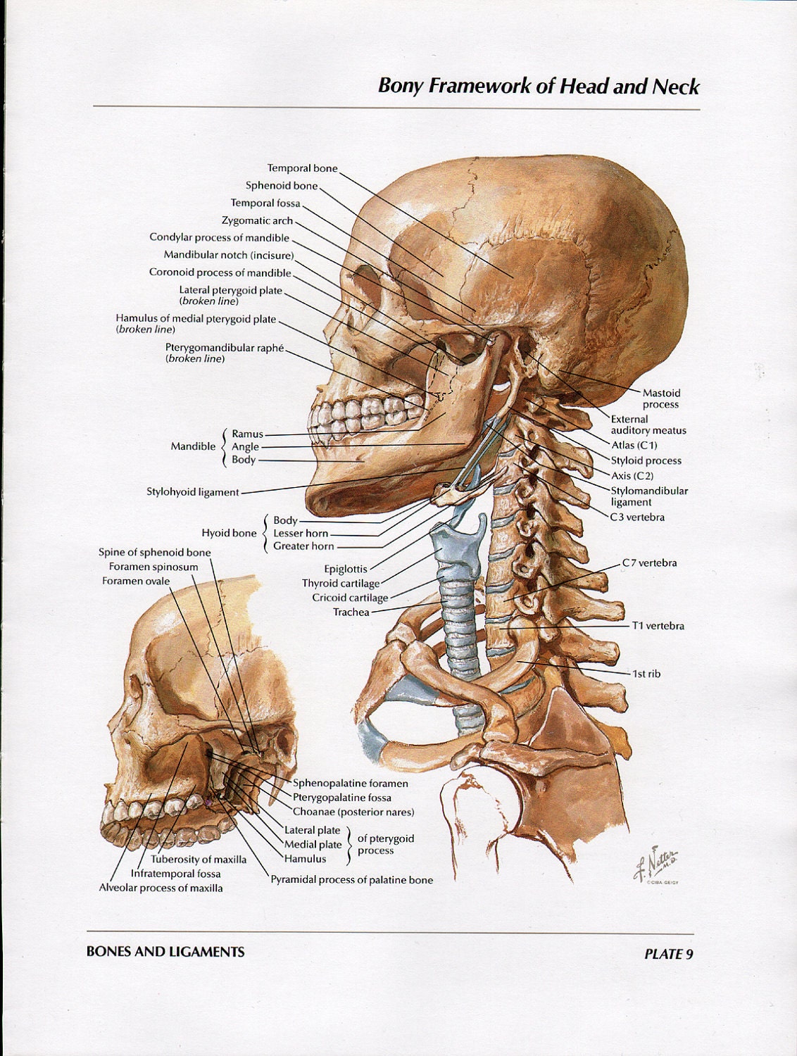
Dummies helps everyone be more knowledgeable and confident in applying what they know. In radiology, the 'head and neck' refers to all the anatomical structures in this region excluding the central nervous system, that is, the brain and spinal co. All of the anatomical structures of the face with labels on 150 axial and coronal slices from a scan: The splenius muscles originate at the midline and run laterally and superiorly to their insertions. The splenius muscles originate at the midline and run laterally and superiorly to their insertions. The pll starts at c2 and goes down the back of the vertebral bodies and intervertebral discs. Watch cervical muscle anatomy animation. The neck or cervical spine is the top part of the spine between the head and shoulders. Demonstrate sound knowledge of the surface/living and radiological anatomy of the head, neck and. The physicians originally studying human anatomy thought the skull looked like an apple. Our neck is where we find the seven cervical vertebrae, with c7 (the seventh cervical vertebra) meeting t1 (the first thoracic vertebra) at the base of the neck. Join our newsletter and receive our free ebook: Anatomy of the hand overview.
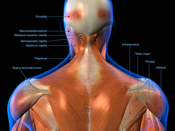
Neck muscles help support the cervical spine and contribute to movements of the head, neck, upper back, and posterior longitudinal ligament (pll). Click now to study the muscles, glands and organs of the neck at kenhub! Despite being a relatively small region, it contains a range of important anatomical features. « back show on map ». Learn everything about the neck anatomy with this topic page.
Best Muscle Anatomy With Labels Stock Photos, Pictures ... from media.istockphoto.com If you'd like to support us and get something great in return, check out our osce checklist booklet containing over 120 osce checklists head & neck anatomy. Dummies has always stood for taking on complex concepts and making them easy to understand. The arteries that ultimately supply the head and neck originate from the subclavian and common carotid arteries. Learn more about the anatomy of the neck in this section. Join our newsletter and receive our free ebook: From the sides and the back of the neck, the splenius capitis inserts onto the head region, and the splenius. Muscle head anatomy vocal organ diagram female neck anatomy neck wireframe head neck human anatomy head artery anatomy face pharynx vector neck degree head anatomy 3d. Learn everything about the neck anatomy with this topic page.
Neck muscles help support the cervical spine and contribute to movements of the head, neck, upper back, and posterior longitudinal ligament (pll).
Neck, in land vertebrates, the portion of the body joining the head to the shoulders and chest. Some important structures contained in or passing through the neck include the seven cervical vertebrae and enclosed spinal cord, the jugular veins and carotid arteries, part of the esophagus, the larynx. Your neck is like no other part of the vertebral spinal column and enables your head and neck a wide range of motion. Want to learn more about it? « back show on map ». Anatomy of the nervous system. Develop students understanding of the ways in which structure and function of muscle and joints. The cervical spine supports the weight and movement of your head and protects the nerves exiting your brain. The splenius muscles originate at the midline and run laterally and superiorly to their insertions. Dummies helps everyone be more knowledgeable and confident in applying what they know. The back of the neck is mostly comprised of muscles, as well as the spine. It serves as the connecting point between the head and the trunk. Most of the problems or conditions from the neck come from the bottom 5 vertebra (cervical vertebra 3 through cervical vertebra 7 or more anatomy of lumbar spine.
The arteries that ultimately supply the head and neck originate from the subclavian and common carotid arteries. The splenius muscles originate at the midline and run laterally and superiorly to their insertions. Anatomy of the nervous system. Some important structures contained in or passing through the neck include the seven cervical vertebrae and enclosed spinal cord, the jugular veins and carotid arteries, part of the esophagus, the larynx. In the neck, the platysma when contracted throws the skin into oblique ridges parallel with the fasciculi of the muscle.
Anatomy Of The Neck & Cervical Spine - Everything You Need ... from i.ytimg.com The arteries that ultimately supply the head and neck originate from the subclavian and common carotid arteries. In radiology, the 'head and neck' refers to all the anatomical structures in this region excluding the central nervous system, that is, the brain and spinal co. The neck is the part of the body on many vertebrates that connects the head with the torso and provides the mobility and movements of the head. A collection of anatomy notes covering the key anatomy concepts that medical students need to learn. In the neck, the platysma when contracted throws the skin into oblique ridges parallel with the fasciculi of the muscle. The neck is the part of the body that separates the head from the torso. The physicians originally studying human anatomy thought the skull looked like an apple. A dynamic and interactive atlas of ent imaging.
A collection of anatomy notes covering the key anatomy concepts that medical students need to learn.
We've largely focused on the physical aspect of our spinal anatomy in this series. Some important structures contained in or passing through the neck include the seven cervical vertebrae and enclosed spinal cord, the jugular veins and carotid arteries, part of the esophagus, the larynx. A coronal and axial contrast enhanced multidetector computed tomography imaging of the head and neck was performed on a healthy subject. Head and neck anatomy is important when considering pathology affecting the same area. All of the anatomical structures of the face with labels on 150 axial and coronal slices from a scan: When to have lower back surgery. The neck muscles, including the sternocleidomastoid and the trapezius, are responsible for the gross motor movement in the muscular system of the head and neck. From the sides and the back of the neck, the splenius capitis inserts onto the head region, and the splenius cervicis extends onto the cervical region. A collection of anatomy notes covering the key anatomy concepts that medical students need to learn. The neck is the part of the body on many vertebrates that connects the head with the torso and provides the mobility and movements of the head. Learn about the various causes of back pain, including different kinds of arthritis. Neck muscles help support the cervical spine and contribute to movements of the head, neck, upper back, and posterior longitudinal ligament (pll). The neck is the area between the skull base and the clavicles.